Characterization of bio functionalized semiconductor Nanocrystals or Gold Nanoparticles


The central goal in this research topic is to follow the adaption and incorporation pathways of optically active semiconductor nanocrystal and gold nanoparticle as carriers for biological molecules into cell culture. We functionalize nanoparticles with molecules, which bind specifically to receptors expressed on the surface of cells. It is already known that these molecules get internalized into the cell after binding to their receptor. The internalized ligand often undergoes intralysosomal degradation (figure 1)[1].
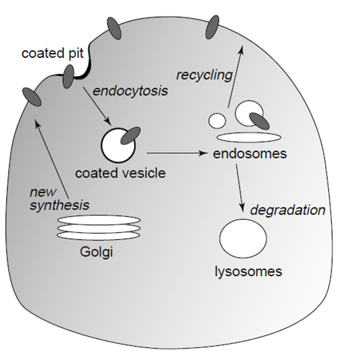
Figure 1: Diagrammatic view of receptor transport pathways [1]
Therefore, we are interested in the investigation of the internalization process by using optically active carriers as transport vehicles for molecules. We use microscopic setups such as Atomic Force Microscopes (AFM) and a Confocal Laser Microscopes (CLM) (figure 2).
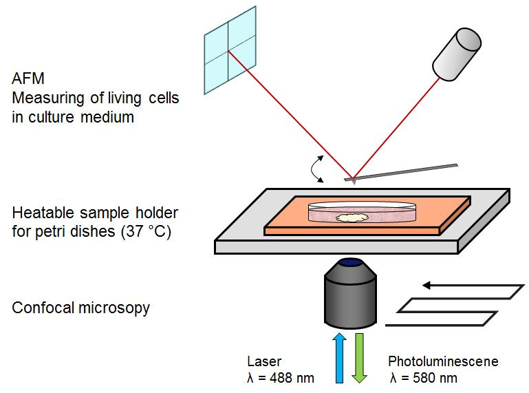
Figure 2: Sketch of the combinatory AFM/CLM setup. A heatable sample holder is necessary for measuring living and healthy cells.
With this combination, we are able to determine the structure and dynamics of cells as well as the position of the functionalized nanoparticles. Hence, the influence of nanoparticles covered with biological active molecules to the cells can be studied (figure 3).
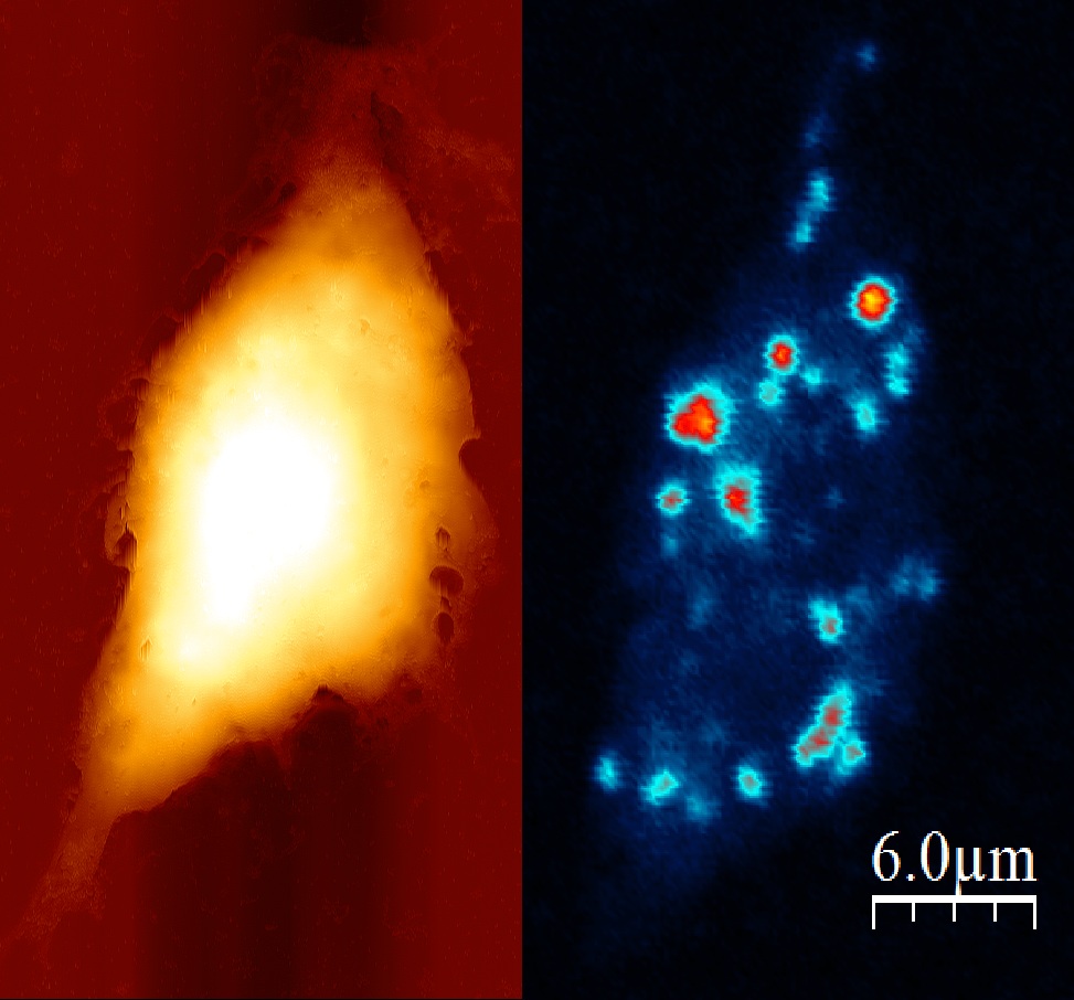
Figure 3: Left image eukaryotic cell measured by AFM in liquid,
right image eukaryotic cell measured by CLM (lighter spots shows the autofluorescence of the cell).
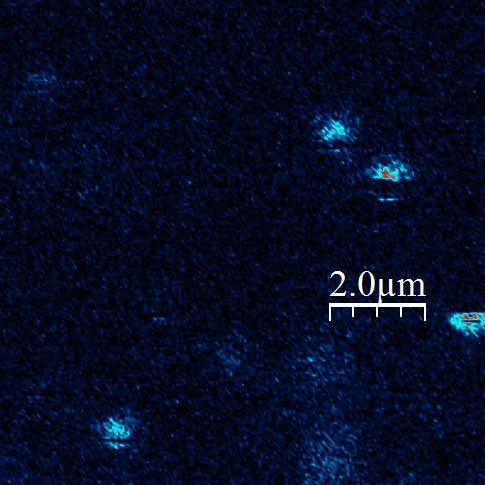
Figure 5: Single CdSe/CdS/ZnS Quantum Dots with Carboxygroup on the surface in confocal scan.
The Scan (left) and also the Video (right) shows on and off times of the fluorescence (“blinking”).
[1] Koenig, JA, Edwardson, JM.: Endocytosis and recycling of G protein-coupled receptors. Trends Pharmacol Sci. 1997 Aug; 18(8):276-87.